

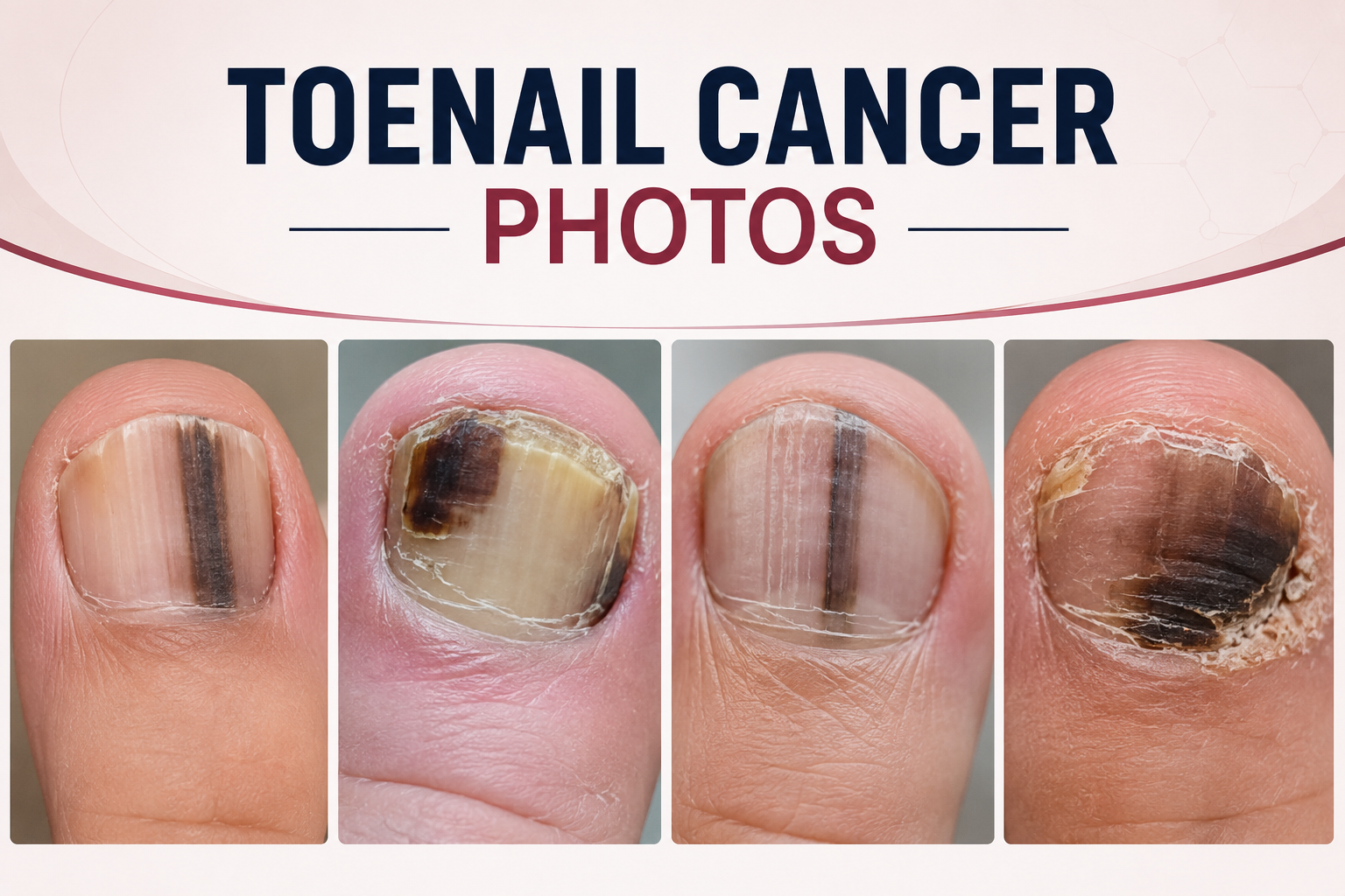
A visual guide to suspicious nail changes, common look-alikes, and when to see a dermatologist
A dark line, brown patch, split nail, or unexplained growth under a toenail can be alarming. Most nail discoloration is caused by injury, fungal infection, pressure from footwear, or normal pigment variation. However, melanoma can rarely begin in the nail unit, and early changes are sometimes mistaken for a bruise or fungus.
People searching for toenail cancer photos are usually trying to compare a new mark with examples online. Pictures may help you notice a pattern, but they cannot confirm whether a streak is cancer. A new, widening, irregular, or persistent change deserves an in-person examination, especially when it affects one nail and does not move outward as the nail grows.
| KEY TAKEAWAYS
· Nail melanoma often appears as a brown or black vertical band, but some cases are red, pink, flesh-colored, or nearly colorless. · A concerning band may widen near the cuticle, contain uneven colors, blur at the edges, or spread pigment onto the surrounding skin. · Toenail fungus more often causes yellow-white discoloration, thickening, crumbling, debris, and involvement of more than one nail. · A bruise usually follows trauma and gradually moves toward the nail tip as the nail grows. · A dermatologist may use dermoscopy, but a nail-unit biopsy is the test that can confirm or rule out melanoma. |
What Does Toenail Cancer Usually Mean?
The phrase “toenail cancer” usually refers to melanoma arising in the nail unit, often called nail melanoma or subungual melanoma. It develops from pigment-producing cells in the skin beneath or around the nail rather than from the hard nail plate itself. The big toe is a common location, although any toenail or fingernail can be affected.
Nail melanoma is uncommon, but delayed diagnosis can make treatment more complicated. It is not usually linked to ultraviolet exposure in the same way as many skin melanomas. It can occur in any skin tone, and nail changes deserve attention in people with darker skin because acral and nail-area melanomas represent a larger share of melanomas diagnosed in these populations.
What Toenail Cancer May Look Like in Photos
Online images show a wide range of appearances. A single photograph is not a diagnostic checklist, because lighting, polish residue, nail thickness, skin tone, camera settings, and previous injury can change how a nail looks. The most useful question is not whether your nail perfectly matches one picture, but whether it has a concerning pattern and is changing over time.
| Photo feature | What it may look like | Why it matters |
| Vertical brown or black band | A stripe running from the nail base toward the tip | More concerning if new in adulthood, widening, irregular, or limited to one nail |
| Wider pigment near the cuticle | A triangular band that broadens at the base | Growth near the nail matrix can make the band wider where it begins |
| Pigment on nearby skin | Brown or black color extending onto the cuticle or side nail fold | This may be called Hutchinson sign and needs prompt assessment |
| Nail splitting or distortion | Cracking, thinning, lifting, or loss of normal shape | Persistent structural change can occur with melanoma, trauma, infection, or psoriasis |
| Bleeding or a nonhealing area | Recurrent blood, crust, ulceration, or a bump under the nail | A lesion that repeatedly bleeds or does not heal should be examined |
| Pink or flesh-colored growth | A pale, red, or colorless bump with little visible pigment | Some nail melanomas lack dark pigment and can resemble a wart or inflamed tissue |
The Nail Melanoma Warning Signs Dermatologists Emphasize
The American Academy of Dermatology advises checking for a new dark streak, a band that changes, darkening of the skin next to the nail, nail lifting, splitting, or a bump beneath the nail. Its nail melanoma photo guide also shows why color extending beyond the nail plate should not be ignored.
Features that raise concern include a stripe wider than nearby benign lines, multiple shades of brown or black, fuzzy borders, rapid widening, new onset in one nail, pigment that reaches the surrounding skin, and nail damage without a clear injury. Persistent pain, swelling, bleeding, or a mass beneath the nail also warrants evaluation, even if the lesion is not dark.
Toenail Melanoma vs. Fungus vs. Bruise
Fungal nails and old bruises are far more common than nail melanoma, but visual overlap is real. The comparison below can help organize what you notice, not diagnose it.
| Feature | Possible nail melanoma | Fungal nail infection | Bruise under the nail |
| Typical color | Brown or black band; sometimes red, pink, or colorless | White, yellow, tan, or brown | Red-purple, dark brown, or black |
| Pattern | Often a lengthwise band or focused lesion | Diffuse discoloration, side streaking, or patchy change | A blotch beneath the nail after impact or pressure |
| Nail texture | May split, lift, thin, distort, or develop a mass | Often thick, brittle, crumbly, and filled with debris | Usually normal except when trauma damaged the nail |
| Number of nails | Often one nail | May spread to several toenails | Usually the injured nail only |
| Movement with growth | Pigment may persist at the nail base or continue widening | Changes persist until infection is treated and healthy nail grows | The dark area generally moves toward the tip with nail growth |
| Helpful next step | Prompt dermatologist examination; biopsy if suspicious | Clinical exam and nail testing before long treatment | Monitor growth if injury is clear; seek care for severe pain or uncertainty |
What Toenail Fungus Usually Looks Like
Fungal nail infection commonly makes a toenail thick, fragile, cracked, discolored, or separated from the nail bed. It may begin at an outer edge and gradually spread, and athlete’s foot may be present between the toes. The CDC overview of fungal nail infection notes that infected nails can change color, become thick, and break easily. Because psoriasis, trauma, and melanoma can mimic fungus, testing is often sensible before committing to months of antifungal treatment.
What a Toenail Bruise Usually Looks Like
A subungual hematoma is trapped blood beneath the nail after stubbing a toe, dropping an object on the foot, running in tight shoes, or repeated athletic impact. It can look red, purple, brown, or black. A bruise generally has a believable injury story and slowly travels toward the nail tip as new nail grows from the base. Toenails grow slowly, so complete clearing can take many months.
Seek urgent care after major trauma if the toe is deformed, pain is severe, bleeding will not stop, or you may have a fracture. A dark area with no remembered injury, one that remains connected to the cuticle, or one that widens instead of growing out should be checked.
Other Conditions That Can Resemble Toenail Cancer
Benign melanonychia. Normal pigment can create one or more lengthwise bands, especially in people with darker skin. Stable, uniform lines involving several nails are often less concerning, but a dermatologist should assess a new or changing band.
Nail psoriasis. Pitting, lifting, thickening, discoloration, and small blood streaks may affect the nails. Skin or joint symptoms can provide additional clues.
Bacterial infection. Green, yellow, red, or dark discoloration can occur with infection, often with tenderness, swelling, drainage, or odor.
Warts or benign growths. A rough bump can distort the nail or resemble a colorless melanoma.
Medication or systemic illness. Some drugs and health conditions can alter nail color, shape, or growth across multiple nails.
Some autoimmune and inflammatory illnesses also produce distinctive skin or nail changes. A broader visual guide to dermatomyositis rash pictures illustrates why the overall symptom pattern matters more than matching one isolated image.
How Doctors Diagnose a Suspicious Toenail
A dermatologist will ask when the change began, whether it is growing, whether there was trauma, which medications you take, and whether melanoma or unusual nail pigment runs in your family. The clinician examines every nail and the surrounding skin, often using a dermatoscope to magnify pigment patterns.
If the appearance suggests fungus, a clipping or scraping may be examined or cultured. If melanoma cannot be excluded, the key test is a biopsy taken from the nail matrix, nail bed, or suspicious tissue. The procedure requires skill because the location of the sample matters and the nail unit can scar. A biopsy can distinguish melanoma from benign pigment, bleeding, infection, and other growths.
What Treatment May Involve
Treatment depends on what the biopsy finds and how deeply abnormal cells extend. Early melanoma may be removed with surgery that clears the tumor and an appropriate margin. More advanced disease may require wider surgery, evaluation of nearby lymph nodes, imaging, or systemic treatment coordinated by dermatology and oncology specialists. The exact operation varies, and modern care does not automatically mean amputation for every nail melanoma.
Fungal nail infection is treated differently and may require prescription topical or oral antifungal medicine after testing. A bruise usually grows out on its own unless pressure, pain, or associated injury requires treatment. These very different pathways are why self-diagnosis from photos can delay the right care.
When Should You See a Dermatologist?
| ARRANGE PROMPT MEDICAL REVIEW IF YOU NOTICE:
· A new brown or black streak in one nail, particularly if it is widening or changing · Pigment spreading onto the cuticle or skin beside the nail · A nail that splits, lifts, bleeds, becomes painful, or develops a persistent bump without clear trauma · A dark mark that does not move toward the tip as the nail grows · A presumed fungal nail that does not improve as expected or was never tested · Any persistent nail change that worries you, especially with a personal or family history of melanoma |
Repeatedly checking a nail, comparing dozens of images, or photographing it multiple times each day can intensify health anxiety without providing certainty. The discussion of symptom checking in this guide to posterior auricular lymph nodes offers a useful reminder: document a change reasonably, then let a qualified clinician assess it rather than relying on endless online comparison.
How to Photograph a Nail Change for Tracking
A clear series of photos can help a clinician understand whether a mark is stable or evolving. It should support, not replace, an appointment.
- Remove nail polish and clean the nail gently without scraping underneath it.
- Use bright natural light and avoid filters or flash glare.
- Take one full-foot photo and one close-up from directly above.
- Include a ruler or another fixed size reference beside the nail.
- Repeat no more often than every two to four weeks unless a clinician recommends otherwise.
- Write down pain, bleeding, trauma, new medications, and whether the mark is moving with nail growth.
Frequently Asked Questions
Are all black lines in toenails cancer?
No. Benign pigment, injury, medication effects, and infection can all create dark lines or patches. A new or changing line, particularly in one nail, should be evaluated rather than diagnosed from color alone.
Can toenail melanoma look like fungus?
Yes. Both can cause discoloration and nail distortion. Fungus more often produces thickening, crumbling, debris, and changes in several nails, while melanoma may create an irregular lengthwise band or a focused lesion. Testing may be needed.
Does toenail melanoma always have a black stripe?
No. It may be brown, black, red, pink, flesh-colored, or nearly colorless. Pain, bleeding, splitting, lifting, or a growing bump can be important even without dark pigment.
How can I tell whether a dark spot is growing out?
Photograph the nail with a ruler and compare its distance from the cuticle over time. A bruise normally moves toward the tip as the nail grows. Pigment that remains at the base or widens needs medical review.
Can a pedicure cause a dark toenail?
Pressure, cutting, friction, or minor trauma during nail care can cause bleeding or inflammation. However, do not assume a persistent or changing mark is from a pedicure if the timing and growth pattern do not fit.
Who should check a suspicious toenail?
A board-certified dermatologist is the most appropriate specialist. A podiatrist or primary care clinician can also examine the toe and refer you when a biopsy or specialized nail assessment is needed.
Should I try antifungal treatment first?
Not automatically. If the diagnosis is uncertain, ask about nail testing before beginning a long treatment course. Treating an unconfirmed “fungus” for months can postpone evaluation of another condition.
The Bottom Line
Photos can teach you which nail changes deserve attention, but they cannot reliably separate melanoma from fungus, bruising, benign pigment, or inflammatory nail disease. The most concerning patterns are new, irregular, widening, persistent, or destructive changes in one nail, especially when color extends onto nearby skin. Early evaluation is the safest next step, and a biopsy can provide the answer when the appearance remains uncertain.
| Medical disclaimer: This article is for general education and does not diagnose nail cancer, fungal infection, or any other condition. Seek professional medical evaluation for a new, changing, painful, bleeding, or persistent nail abnormality. |